


SEM – Scanning Electron Microscopy
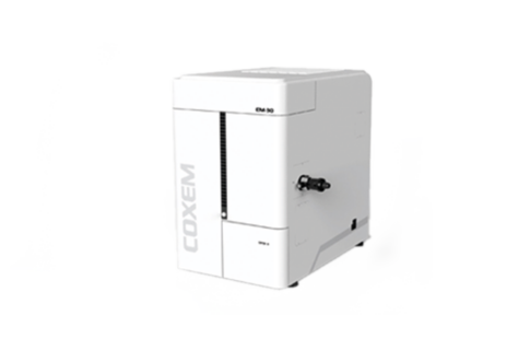
Brand / Model
COXEM EM-30 Plus
Features
Magnification: x20 – x150k (~ x80k)
Accelerating Voltage: 1kV to 30kV
Electron Gun: Tungsten Filament (w)
Detector: SE Detector, BSE Detector
Max. Sample Size: 45mm (H), 30mm (Diameter)
Vacuum System: Turbo molecular pump (less than 3min),
low vacuum mode
Aim
The SEM image is obtained by concentrating the high voltage accelerated electrons in the high vacuum environment on the sample, collecting the products formed as a result of various interactions between the electron and sample atoms during the scanning of this electron beam on the sample surface, and then transferring them to the screen as a result. Thanks to secondary electrons, a high resolution topographic image of the sample is obtained. Furthermore, it is possible to analyze the insulating samples, polymers and glass samples that are not plated with low vacuum mode. It is used in researching the morphology of natural resources, determination of defects and wear properties of materials, determination of properties such as surface properties of materials, coating thickness, particle size, morphology, etc.
Terms of sample delivery:
• Powder samples should be at least 30 mg.
• It should be noted that the samples are conductive or insulating.
• Samples should not contain moisture.
• Solid sample and thin film sample sizes should be max 3×3 cm and height should be max 2 cm.
Click here for application